We are proud to announce that

the 16th Annual EFCS Tutorial

Halle, Germany, 15/09/2025 – 19/09/2025

organized by European Federation of Cytology Societies

has been accredited

by the European Accreditation Council for Continuing Medical Education (EACCME®)

for a maximum of 31.5 European CME credits (ECMEC®s).

CYTOPATHOLOGY JOURNAL BECAME AN OFFICIAL JOURNAL OF THE EFCS

It was a final result of a long lasting, but fruitful conversation with Wiley, the journal publisher, British Association for Cytopathology (BAC) and EFCS. BAC was Affiliated Society of Cytopathology Journal for decades, and in the future, being also an EFCS member, its engagement will continue inside the wide family of European Cytology Societies. We wish to thank representatives of the BAC which played a major role in achieving this friendly agreement.

Editorial Board of Cytopathology Journal is a team of renowned European and International experts led by Philippe Vielh (Editor-in-Chief) and Ashish Chandra (Deputy Editor).

We invite you to read the Cytopathology Journal, as well as considering a manuscript submission of your research and professional work.

The link for the journal: https://onlinelibrary.wiley.com/journal/13652303

Case of the Month by Morgane Dubois

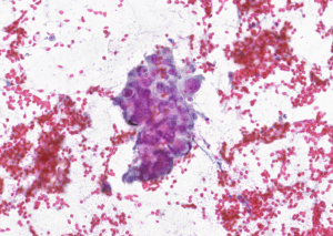
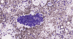
CLINICAL PRESENTATIONThis is a 77-year-old woman with a 10-mm BI-RADS 4 mass in the upper inner quadrant ofthe left breast. Core needle biopsy shows invasive carcinoma of no special type (NST),Nottingham grade II (Elston–Ellis). Hormone receptors positive (100%), HER2 1+,

Read More »

16th annual EFCS cytopathology tutorial report

The 16th annual EFCS cytopathology tutorial took place in Halle, Germany, from 15 to 19 September 2025. Giovanni Negri chaired the event, while Maurizio Pinamonti was the co-chair. Ralf Heine was the local host.  Following the usual format, the tutorial

Read More »

Case of the Month by Mario Urso

On 4 March, a 49-year-old man was referred to the Fine Needle Aspiration (FNA) Clinic at our hospital for evaluation of a large thyroid nodule. Neck ultrasound revealed a normally positioned thyroid gland with a 6 cm hypoechoic nodular mass

Read More »